English


-
Machinery
Choose from 20,092 used machinery listings
DESCRIPTION
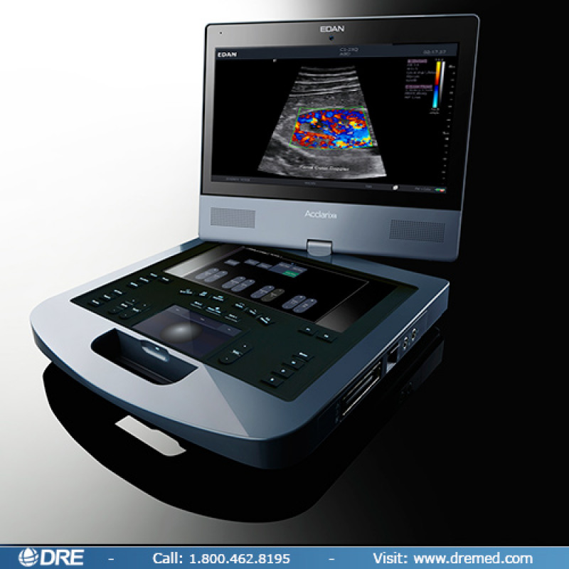
The remarkable Edan Acclarix AX8 Portable Ultrasound System delivers a powerhouse of features in an exceptionally mobile package.
The Acclarix AX8 ultrasound system is a fully featured portable diagnostic ultrasound platform designed from the ground up with a relentless focus on delivering unexpected levels of innovation and performance. Born of a vision to deliver meaningful innovations that solve real clinical challenges
the Acclarix AX8 features a distinctive design, definitive image quality, intelligent workflow and intrinsic quality.
Distinctive Design
Sleek, compact design facilitates portability for use in any care environment.
15" HD LCD monitor tilts and swivels for optimal viewing during procedures.
Fully sealed control panel, aids in maintaining infection control.
Removable lithium ion battery with dual touch sensors to rapidly check battery level.
Definitive Image Quality
A comprehensive suite of transducers cover a variety of applications and provide excellent detail, contrast resolution and image uniformity.
Standard system configuration includes advanced image optimization technologies, including speckle reduction and spatial compounding technologies.
High fidelity, high channel count system architecture.
Tissue Adaptive Imaging is a unique core technology available in all imaging modes. It continuously and automatically optimizes for the anatomy being scanned to provide exceptional image quality and consistency to reduce the need for user optimization.
Intelligent Workflow
Dual, gesture-control touch screens streamline workflow for easy operation.
Customizable 10” touch screen allows prioritization of functions to personalize workflow.
B-mode Auto optimize for one touch gain optimization.
Doppler Flow quick-sets provide one touch flow optimization.
Needle visualization feature enhances the ability to identify the needle quickly, even while encountering steep angles.
Multi-transducer connect option allows three transducers to be connected simultaneously so the transducer you need is always available.
Large capacity 500 GB hard drive, 4 USB ports and DICOM connectivity for flexibility in archiving images.
Intrinsic Quality
Designed with high quality materials for durability.
Highly responsive touch screens, which maintain sensitivity even while wearing gloves.
User interface hard keys are sealed with silicon rubber for resiliency and superb tactile feedback.
15” monitor with specialized, bonded glass for durability and enhanced image clarity.
Ultrasound Image Application Library
Anesthesia
General Imaging
MSK - Orthopedic
OB GYN
Highly Flexible Across Multiple Applications
The versatile Acclarix AX8 is ideally suited for a variety of applications, procedure guidance and clinical specialties including:
Regional Anesthesia
Pain Management
Musculoskeletal
Rheumatology
Sports Medicine
Orthopedic Surgery
Physical and Rehabilitation Medicine
Obstetrics and Gynecology
General Imaging
Interventional Radiology
Available Transducers
C5-2XQ
Format: Curved linear array
Footprint: 60mm
Fundamental Frequencies: 2.0-5.0 MHz Curved linear array
Harmonic Frequencies: H3.0-5.0 MHz
Supported Applications: Spine, Abdomen, OB, Gynecology
L10-4Q
Format: Linear array
Footprint: 38mm
Fundamental Frequencies: 4.0-9.0 MHz
Harmonic Frequencies: H6.0-10.0 MHz
Supported Applications: Small parts, MSK, Nerve, Vascular
L17-7HQ
Format: High frequency linear
Footprint: 38mm
Fundamental Frequencies: 7.0-14.0 MHz
Harmonic Frequencies: H9.0-17.0 MHz
Supported Applications: Small parts, MSK, Nerve, Vascular
E8-4Q
Format: Endocavity tightly curved array
Footprint: 10mm
Fundamental Frequencies: 4.0-8.0 MHz
Harmonic Frequencies: H5.0-8.0 MHz
Supported Applications: OB, Gynecology, Prostate, Endovaginal, Endorectal
P5-1XQ
Format: Phased array
Footprint: 21mm
Fundamental Frequencies: 1.0-5.0 MHz
Harmonic Frequencies: H2.0-5.0 MHz
Supported Applications: Adult Cardiac, Abdomen ( including difficult to image )
L17-7SQ
Format: High-frequency compact linear array
Footprint: 20mm
Fundamental Frequencies: 7.0-14.0 MHz
Harmonic Frequencies: H9.0-17.0 MHz
Supported Applications: MSK, Nerve, Vascular, Intraoperative
SPECIFICATIONS
System Architecture
128 channels
Quad beam
i7 processor with quad virtual cores
16Gb memory
500Gb hard drive storage
Mechanical Packaging
Dimensions: W 38.8cm, D 40.7cm, H 7.7cm
Weight: 9.25kg (includes battery)
Main Screen: Tilt- and-swivel adjustment | 15” HD resolution (1920x1080)
Magnetic monitor latch for secure transport
Integrated handle provides wrist support during imaging
Removable battery provides approximately 60 minutes of typical ultrasound use
Optional multi-transducer connector
Durable, ergonomic carry case
B-mode Imaging
Tissue Adaptive Imaging provides continuous and automatic optimization including: dynamic range, speckle reduction, spatial compounding and persistence
Enhanced border detection algorithms
One-key auto optimization
Digital zoom
Depth: up to 30cm ( transducer dependent )
Frequency range: Up to three fundamental and two harmonic frequencies per transducer
Frequency compounding enhances penetration and detail
Spatial compounding
Speckle reduction
Imaging formats: Curved, Linear, Phased array, Trapezoid, FOV for increased frame rate, Up/Down, Left/Right invert, Linear steered, Dual
Additional optimization parameters: Gain and TGC, Dynamic range: 40-96, Map, Tint, Persistent, Focus position and number, Frame rate
Color Doppler
Adaptive Doppler Imaging Automatically and continuously adapts to the flow state to optimize color fill-in, boundary detection and hemodynamic display.
Supported modes: Velocity, Power Doppler Imaging (PDI), Directional PDI (DPDI)
Side-by-side live format B-mode/color Doppler
Additional optimization parameters: Gain, dynamic range, frame rate, frequency | Persistence, smoothing, wall filter, map | Steer angle, scale, invert, baseline, threshold
Strip Doppler
Supported modes: PW, CW
HPRF: Automatic invocation as needed to maintain gate location/scale
Auto Doppler measurements: User selectable sensitivity and direction
Duplex and Triplex displays
Additional optimization parameters: Scale, Gain, Dynamic range, Wall Filter, Sweep speed, Baseline, Angle, Steer, Invert, Volume, Map and Tint, Frequency, Gate size, Strip size: Selectable top-bottom split screen display including full strip
M-mode
Optimization parameters: Sweep speed, Persist, Map and tint, Strip size: Selectable top-bottom split screen display including full or side-by-side format, Gain, frequency, dynamic range (shared with B-mode)
Advanced Features
Tissue Adaptive Imaging
Adaptive Doppler Imaging
Needle visualization improves identification, even at steep angles
Panorama
Frequency compounding
Spatial compounding
Speckle reduction
B-mode auto-optimization
User Interface
Touch screen has three levels of access and drag-and-drop functionality for quick customization: Core functionality on one page, Swipe between pages for second tier controls, User created folders store infrequently used controls
Unique dual touchscreen with second screen housing electronic virtual trackball: Gesture driven UI, Swipe to change, gain, scroll cine, Pinch in/out to zoom and resize color ROI
Hard keys to access to core controls: Provides tactile feedback and landmarks for eyes-up navigation
Sealed for easy cleaning
Two programmable hard keys for direct access to most frequently used features
Languages: English, Spanish, Ask about additional options
Environmental Operating Requirements
Ambient temperature: 0° to 40°C
Relative Humidity: 20%~80% (no condensation)
Atmospheric pressure: 700hPa-1060hPa
110V-240V power supply
Cart Specifications (Optional)
Snap-in anchoring of laptop into cart
Height range of 31” to 40” to laptop palm rest
Fixed deck angle of 15°
Power converter housing built under tray
Built-in tray houses printer and other incidentals
Connectivity
DICOM: Verify SCP, Static image store SCU, Ultrasound multi-image store SCU, Four levels of compression, Data transfer options, Removable media, In-progress network storage, Auto-store at exam end, Manual-store on demand
4 USB ports (2 USB 2.0; 2 USB 3.0) Exports: DICOM studies, AVI and BMP files, PDF of report
Video out: Display port and S-video
Ethernet
This equipment is located in US
| Manufacturer | Edan Acclarix A |
| Model | Edan Acclarix A |
| Year | 2017 |
| Country |
 USA
USA
|
| Condition | Good |
| Main category | Lab, Medical and Bioscience |
| Subcategory | Medical Equipment |
| ID | P81109055 |
| Client type | Machinery dealer |
| On Kitmondo since | 2018 |
| Number of listings | 0 |
| Country |
USA
|
| Last activity | Nov. 21, 2018 |
 Germany
 Czechia

 Cyprus
 Italy
Italy